Jessica Kidd Equine Surgeon
  • Home
  • About
  • Services
  • Affiliated clinics and hospitals
  • Referral process
  • Links
  • Case Studies
  • Contact
  • News
  • Home
  • About
  • Services
  • Affiliated clinics and hospitals
  • Referral process
  • Links
  • Case Studies
  • Contact
  • News
Search by typing & pressing enter

YOUR CART

Case studies

These are a few recent cases who underwent surgery with Jessica Kidd. More to follow!
Foal with carpal valgus deformity
Surgery on foal with carpal valgus deformity
Radiograph of foal with carpal valgus deformity
A three month old foal with a "bent knee". This foal had an abnormal angle to the right knee, which is termed a "carpal valgus deformity". The knee was not correcting on its own and the age of the foal meant a limited window of opportunity for surgical correction. The foal underwent surgery for placement of a transphyseal screw, meaning a screw which crosses the growth plate, and aims to slow the growth on one side of the knee and allow the slower side to "catch up".  Once the knee is straight, the screw will be removed.


A show pony with a wound to her nose. The pony had lacerated her nostril and the wound did not heal well the first time. She was admitted for revision of the wound and reconstruction of her nose. This case was carried out in conjunction with Georgie Hollis at The Vet Wound Library (http://www.vetwoundlibrary.com/ and see links) which is an excellent resource for wound cases.

Horse nose laceration
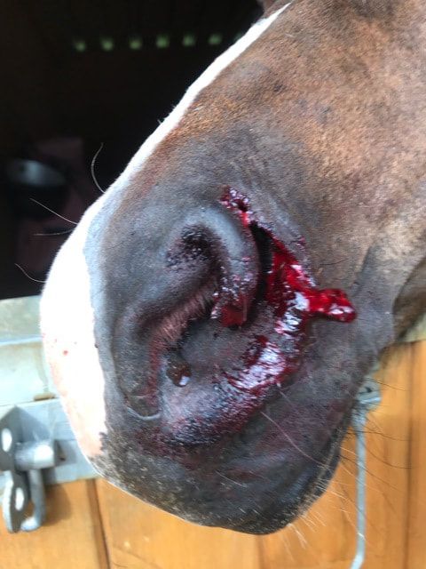
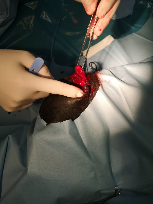
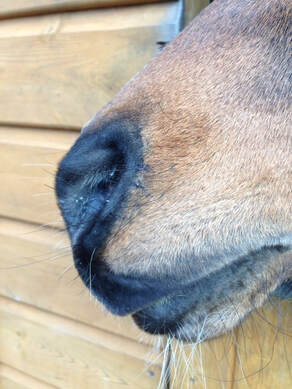
The mare just after she caught her nose on a nail.
Horse nose laceration poor healing
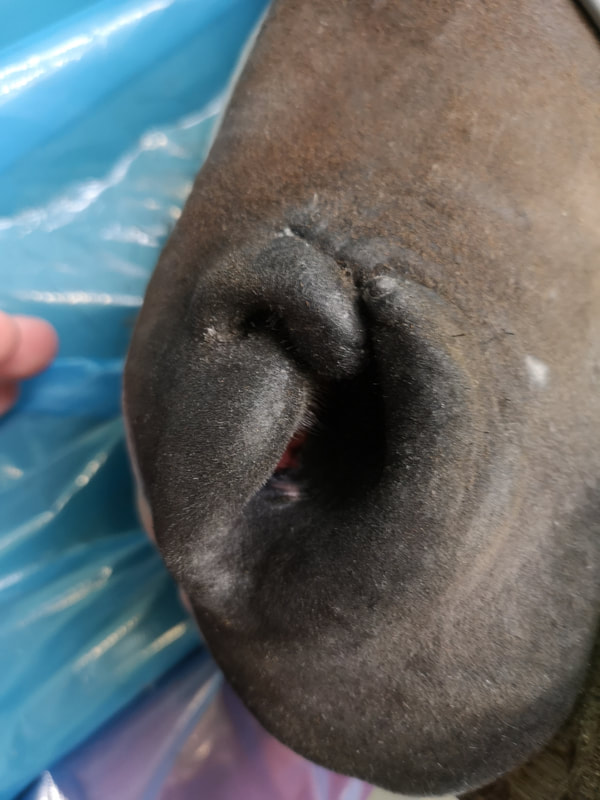
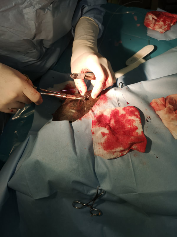
The nose as it had initially healed, just before the surgical revision.
Horse nose laceration healing revision surgery
Horse nose laceration after revision surgery
Result after revision surgery
The mare this week.


 This mare went to a new owner who was concerned about the melanomas under her tail and in her mouth.
The entrenched veterinary view is that melanomas are benign and should be left alone. What we know now, from research by Professor Derek Knottenbelt, is that external melanomas (those we can see on a horse), if left untreated, will in time develop into internal (cannot be seen and involving internal organs) melanomas, for which we have no effective treatments. And will untimately shorten the horses life span. So I recommend removal of accessible melanomas. Some areas are more difficult than others, in particular melanomas in the throat latch area, but many can be removed entirely. This mare is now back to a happy life and is showing successfully. For much more information, please see the Equine Medical Solutions website link to melanomas: equinesarcoid.co.uk/melanoma-in-horses
Mare melanoma under tail around anus
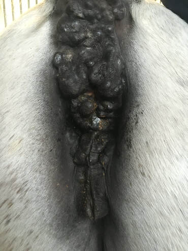
The mare immediately before surgery with extensive melanomas around her rectum and the top of her vulva.
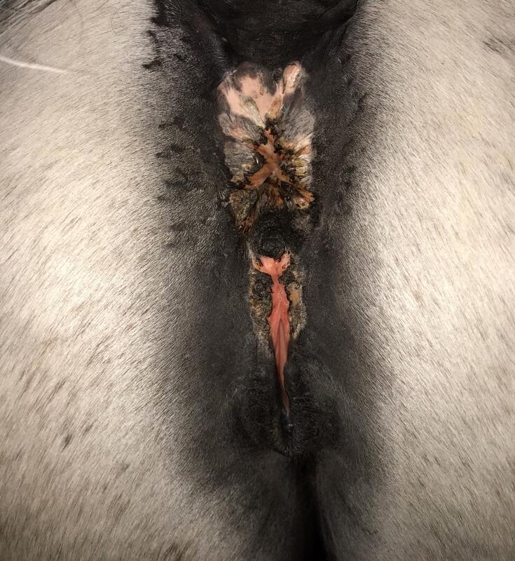
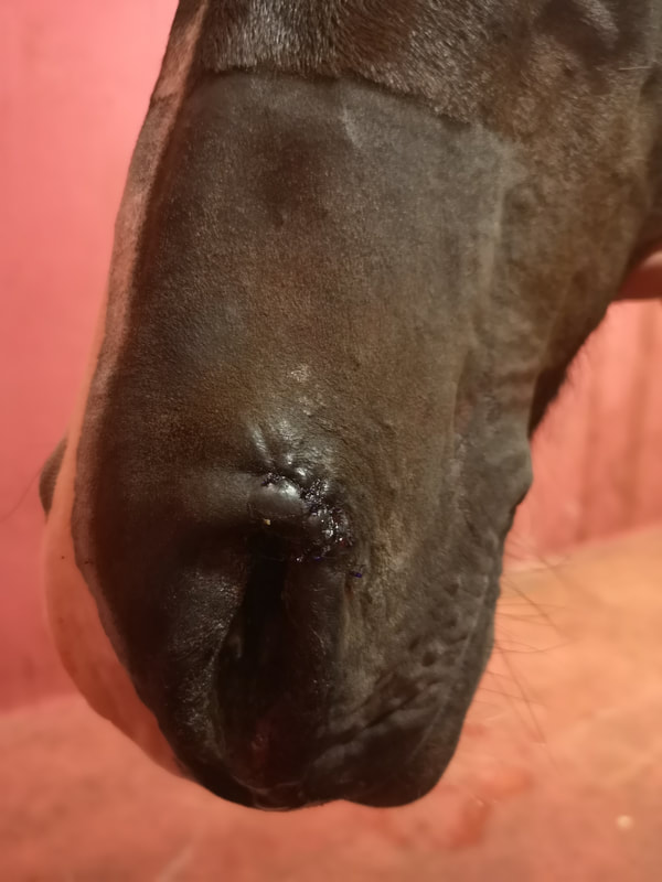
The mare after removal of the melanomas around the anus
The mare currently.


This retired showjumper was presented via his farrier who was concerned over recurrent foot abscesses and an abnormal white line on the sole of the foot. This prompted radiographs which showed two areas in the pedal bone which had been "eaten away" by pressure from an adjacent structure. The radiographs were suggestive of a benign growth of keratin called a "keratoma", but what was very unusual was that there seemed to be two. An MRI was performed at The University of Bristol's vet school to get and idea of the tissues involved and how large these masses were in three dimesions. Using the information from the MRI, surgery proceeded under general anaesthesia and all abnormal tissue was removed from both sites. The tissues were sent to a pathologist who confirmed them to be keratomas. The outlook for these cases is very good, as long as all of the keratoma(s) is removed at surgery. The horse is now in a hospital plate shoe while the solar surface of his foot heals completely.
Radiograph of keratoma
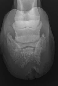
The first xrays which suggested a keratoma (or two).
Picture
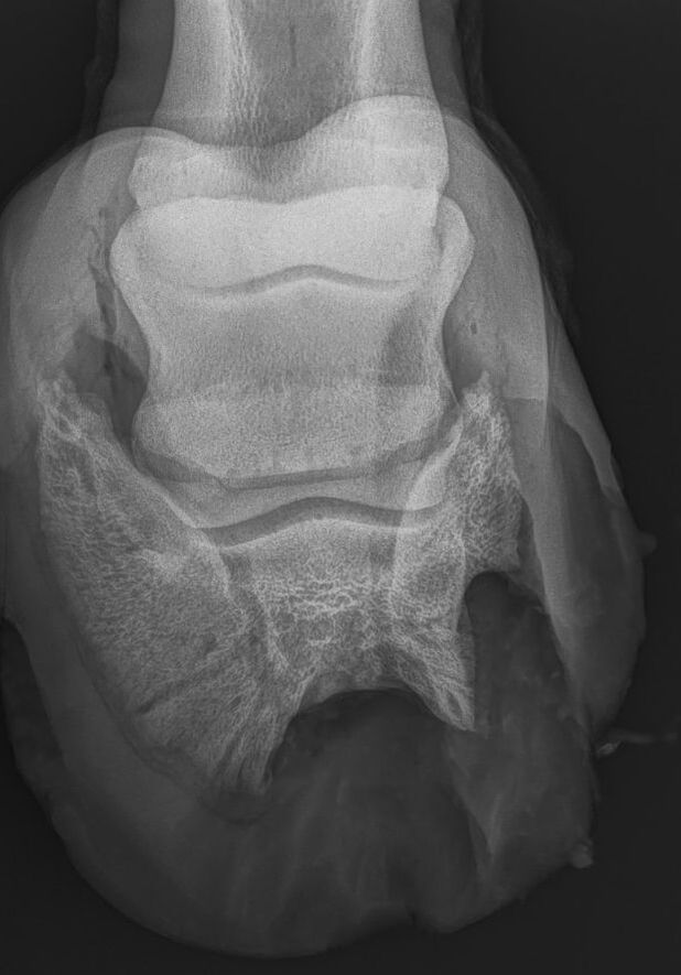
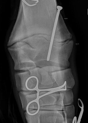
the final radiograph in surgery showing the bone margins are now smooth. Keratomas are not visible on xrays.
Surgery on horse with keratomas
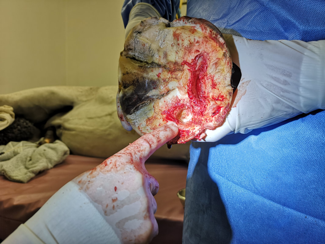
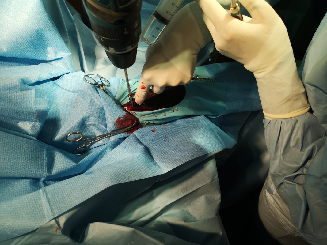
In surgery, my finger is pointing to a small cream coloured mass which is the second keratoma
Hospital plate shoeing for horse with keratoma
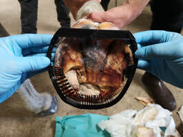
A week after surgery, the farrier Luke Ellis measures the foot for a hospital plate which is a shoes that waterproofs the foot and removes the need for bandaging. The farrier made a "mock up" shoe on his 3D printer!
Home
About
Services
Affiliated clinics and hospitals
Referral Process
Case Studies
Links
Contact
News